43 cell picture and labels
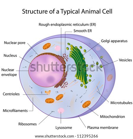
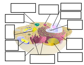
A Labeled Diagram of the Animal Cell and its Organelles A Labeled Diagram of the Animal Cell and its Organelles There are two types of cells - Prokaryotic and Eucaryotic. Eukaryotic cells are larger, more complex, and have evolved more recently than prokaryotes. Where, prokaryotes are just bacteria and archaea, eukaryotes are literally everything else. Human Cell Diagram, Parts, Pictures, Structure and Functions One of the few cells in the human body that lacks almost all organelles are the red blood cells. The main organelles are as follows : cell membrane endoplasmic reticulum Golgi apparatus lysosomes mitochondria nucleus perioxisomes microfilaments and microtubules Diagram of the human cell illustrating the different parts of the cell. Cell Membrane
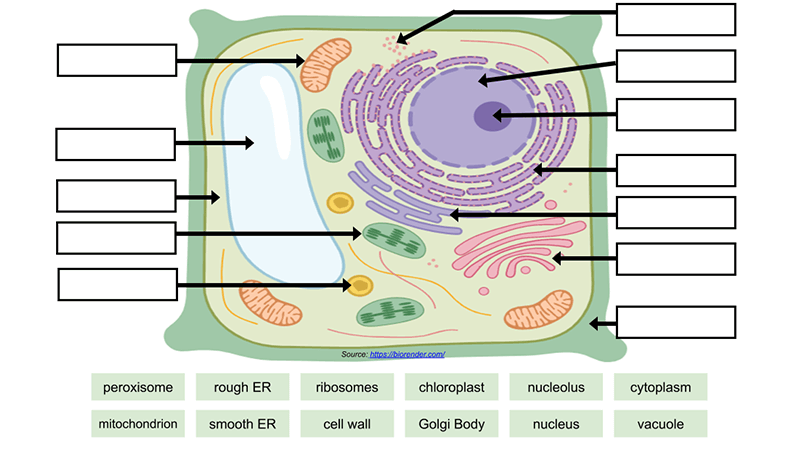
A Labeled Diagram of the Plant Cell and Functions of its Organelles A Labeled Plant Cell Amyloplasts A major component of plants that are starchy in nature, the amyloplasts are organelles that store starch. They are classified as plastids, and are also known as starch grains. They are responsible for the conversion of starch into sugar, that gives energy to the starchy plants and tubers.

Cell picture and labels
Plant and Animal Cell: Labeled Diagram, Structure, Function - Embibe Both plant and animal cells have similar types of architecture. They are made up of cell boundaries, cytoplasm, nucleus and several cellular organelles. Structure. Description and function. Cell Wall. 1. Non-living, rigid, outer boundary. 2. Made up of cellulose, hemicellulose, pectin, lignin, etc. Picture Show: Cell Press The cellular structures featured in this Cell Picture Show are unlikely to appear in general biology textbooks because they are newly described, somewhat rare, and/or their functions are obscure. They range from the astonishing extracellular secretions of algae shown here to subcellular magnets in bacteria and the vaults and "snakes" found in ... Plant Cell Labeled Stock Illustrations - Dreamstime Download 89 Plant Cell Labeled Stock Illustrations, Vectors & Clipart for FREE or amazingly low rates! New users enjoy 60% OFF. 185,513,729 stock photos online.
Cell picture and labels. Describe, with labeled pictures, the cell cycle (interphase, mitosis ... Describe, with labeled pictures, the cell cycle (interphase, mitosis, cytokinesis) STUDY Flashcards Learn Write Spell Test PLAY Match Gravity Created by jiggasmuvPLUS Cell Cycle and mitosis vocabulary Terms in this set (8) Cell cycle A regulated, continuous sequence of preparation (interphase) and division (mitosis) in a cell Interphase Cell Division Stock Photos, Pictures & Royalty-Free Images - iStock Cell Division Pictures, Images and Stock Photos View cell division videos Browse 7,255 cell division stock photos and images available, or search for human cell division or cancer cell division to find more great stock photos and pictures. Newest results human cell division cancer cell division cell division icon cell division abstract Animal Cell Labeled Pictures, Images and Stock Photos Animal Cell Labeled Pictures, Images and Stock Photos View animal cell labeled videos Browse 116 animal cell labeled stock photos and images available, or start a new search to explore more stock photos and images. Newest results Components of Eukaryotic cell, nucleus and organelles and plasma... Diagrams of animal and plant cells Plant and Animal Cells - Labeled Graphics A compilation of plant and animal cell images with organelles and major structures labeled. Students can print images to help them learn the cell. Other Cell Resources. Cheek Cell Lab - observe cheek cells ... if students missed the lab that day they can view a site with pictures to complete lab handout Plant Cell Virtual Lab ...
a picture of a plant cell with labels | plant cell (diagram & label)(7 ... How to Make a Model Cell A cell model is a 3-dimensional structure showing the parts of a plant or an animal cell. You can make a model cell with things from around your house, or you can buy a few simple items to create a fun, educational project. Decide if you... O Orlando Wagner Nats 3d Cell Model Project 3 7th Grade Science Projects Labeled Cell - an overview | ScienceDirect Topics Labeled Cell. The labeled cells show a different color from the background, which makes the cell easy to be segmented. ... The fluorescence microscopy data were based on the photos shown in Fig. 18 A, and the brightness values of the green channel were extracted. The measured data were normalized based on the minimum and maximum cell values. Labelled cell Images, Stock Photos & Vectors | Shutterstock Labelled cell royalty-free images 139,382 labelled cell stock photos, vectors, and illustrations are available royalty-free. See labelled cell stock video clips Image type Orientation Sort by Popular Biology Healthcare and Medical Animals and Wildlife Science Computing Devices and Phones cell anatomy eukaryote bacterium organelle Next of 1,394 03 Label the Cell Diagram | Quizlet Start studying 03 Label the Cell. Learn vocabulary, terms, and more with flashcards, games, and other study tools.
Labeled Plant Cell With Diagrams | Science Trends Photorespiration is the method by which plant cells respirate, taking up light and producing energy. Photorespiration what happens in plants when carbon dioxide levels are too low to produce energy through photosynthesis. The process is somewhat wasteful compared to regular photosynthesis. Cell Organelles- Definition, Structure, Functions, Diagram Cilia and Flagella are tiny hair-like projections from the cell made of microtubules and covered by the plasma membrane. Structure of Cilia and Flagella Cilia are hair-like projections that have a 9+2 arrangement of microtubules with a radial pattern of 9 outer microtubule doublet that surrounds two singlet microtubules. Eukaryotic cell Images, Stock Photos & Vectors | Shutterstock Eukaryotic cell images 4,233 eukaryotic cell stock photos, vectors, and illustrations are available royalty-free. See eukaryotic cell stock video clips Image type Orientation Sort by Popular Biology Animals and Wildlife Healthcare and Medical Science eukaryote cell anatomy nucleus organelle plasma membrane Next of 43 Add graphics to labels - support.microsoft.com Start by creating a New Document of labels. For more info, see Create a sheet of nametags or address labels. Insert a graphic and then select it. Go to Picture Format > Text Wrapping, and select Square. Select X to close. Drag the image into position within the label. and type your text. Save or print your label.
Labeled Diagram Of The Neuron, Nerve Cell That Is The Main Part Of The ... Royalty Free Cliparts, Vectors, And Stock Illustration. Image 48129376. Get 10 FREE Images when you get started on our 1 Month-Free Trial. START NOW Vector — Labeled diagram of the neuron, nerve cell that is the main part of the nervous system. Compare Reset Filter Auto-enhance Background removal Size Standard sizes M 2054 x 2042 px L
Picture Of Prokaryotic Cell With Labels - ClipArt Best Picture of prokaryotic cell with labels You can pick any picture of prokaryotic cell with labels and use it to design a poster to educate medical students about this particular cell. 39 picture of prokaryotic cell with labels .
Telophase Photos and Premium High Res Pictures - Getty Images microscope image of plant cells with three nuclei in anaphase - telophase stock pictures, royalty-free photos & images. whitefish mitosis, whitefish embryo (blastula), telophase and cytokinesis (magnification x400) the cleavage furrow is evident constricting the cell. spindle fibres (microtubules) are still visible. - telophase stock pictures ...
Telophase Labeled Diagram - schematron.org Mitosis: Labeled Diagram. Mitosis is a process of cell Prophase: The chromatin , diffuse in interphase, condenses into chromosomes. Each chromosome has.In telophase II of meiosis, the following events occur: Distinct nuclei form at the opposite poles. Cytokinesis (division of the cytoplasm and the formation of two distinct cells) occurs.
PDF Cell Color, Cut & Paste - Weebly cell, and label them with the correct organelle name. s s) Nucleus Nucleolus Endoplasmic Reticulum (Smooth & Rough) Vacuoel Chloroplast Mitochondria Ribosomes Golgi Bodies Don't Forget: Cell Wall Cell Membrane Cytoplasm. s) s. Plant Cell Answer Key. Plant Cell Color Example % 00 0 00 0 00 0 00 00 Wilh 'he "'red klima' Cell .
Plant Cell Label Images, Stock Photos & Vectors | Shutterstock Plant Cell Label Images, Stock Photos & Vectors | Shutterstock plant cell label images 865 plant cell label stock photos, vectors, and illustrations are available royalty-free. See plant cell label stock video clips of 9
Animal Cell - Free printable to label + Color -kidCourses.com Can you label and color these important parts of the animal cell? NUCLEUS control center for cell (cell growth, cell metabolism, cell reproduction) NUCLEOLUS synthesizes rRNA RIBOSOMES the site of protein building, this is where translation takes place (mRNA in language of nucleic acids is translated into the language of amino acids)
Plant Cell- Definition, Structure, Parts, Functions, Labeled Diagram Functions of the plant cell (plasma) membrane. In-plant cells the cell membrane separated the cytoplasm from the cell wall. It has a selective permeability hence it regulates the contents that move in and out of the cell. It also protects the cell from external damage and provides support and stability to the cell.

452 best images about Cells, Cells, Cells on Pinterest | Cell structure, Cell wall and Science ...
PDF Human Cell Diagram, Parts, Pictures, Structure and Functions One of the few cells in the human body that lacks almost all organelles are the red blood cells. The main organelles are as follows : cell membrane endoplasmic reticulum Golgi apparatus lysosomes mitochondria nucleus perioxisomes microfilaments and microtubules 2
Cell Cycle Labeling | Cell cycle, Biology worksheet, Mitosis Students label the image of a cell undergoing mitosis and answer questions about the cell cycle: interphase, prophase, metaphase, anaphase, and telophase. Find this Pin and more on Education by Indra Sadiq. Biology Classroom Biology Teacher Cell Biology Science Biology Science Education Life Science Ap Biology Forensic Science Teaching Cells
Plant Cell Labeled Stock Illustrations - Dreamstime Download 89 Plant Cell Labeled Stock Illustrations, Vectors & Clipart for FREE or amazingly low rates! New users enjoy 60% OFF. 185,513,729 stock photos online.

Free Beautiful Photos collection: Free Beautiful Cell Phone Wallpapers,Mobile Phone Background ...
Picture Show: Cell Press The cellular structures featured in this Cell Picture Show are unlikely to appear in general biology textbooks because they are newly described, somewhat rare, and/or their functions are obscure. They range from the astonishing extracellular secretions of algae shown here to subcellular magnets in bacteria and the vaults and "snakes" found in ...
Plant and Animal Cell: Labeled Diagram, Structure, Function - Embibe Both plant and animal cells have similar types of architecture. They are made up of cell boundaries, cytoplasm, nucleus and several cellular organelles. Structure. Description and function. Cell Wall. 1. Non-living, rigid, outer boundary. 2. Made up of cellulose, hemicellulose, pectin, lignin, etc.

.jpg)


Post a Comment for "43 cell picture and labels"