43 eye diagram with labels and functions
Eye anatomy and function - AboutKidsHealth A clear lens, located behind the pupil, acts like a camera lens by focusing light onto the retina at the back of the eye. The retina is a light-sensitive inner lining at the back of the eye. Ten different layers of cells work together in the retina to detect light and turn it into electrical impulses. Anatomy of the eye: 3/4 view. Labeled Eye Diagram | Eye anatomy diagram, Eye anatomy ... - Pinterest AS-IS: This vibrant 20" x 26" (55 x 61 cm) exam-room anatomy poster shows cross section of The Eye. It also provides lateral and superior view of the eye and shows the visual field. Anterior chamber angle, eyelashes, tear ducts, cornea, lens, retina, fundus and the macula lutea are illustrated.
Eye Anatomy | Definition, Structure & Functions - iBiologia Diagram of Human Eye with Labelling. Eye Anatomy Complete Physiology of Eye is described below in the given paragraph: The eye is rather like a living Camera. Each eye is a liquid-filled ball 2.5 cm in diameter. At the front of the eye is a clear, round window called the cornea. Behind the cornea is a "lens.

Eye diagram with labels and functions
Anatomy of the eye: Quizzes and diagrams - Kenhub Take a look at the diagram of the eyeball above. Here you can see all of the main structures in this area. Spend some time reviewing the name and location of each one, then try to label the eye yourself - without peeking! - using the eye diagram (blank) below. Unlabeled diagram of the eye. Click below to download our free unlabeled diagram of ... diagram of eye with labelling Eye Diagram With Labels And Functions - Aflam-Neeeak aflam-neeeak.blogspot.com Earthworm Presentation earthworm labeled dissection external anatomy labels presentation lumbricus label section cross slideshare sp fig physiology Labeled Eye Diagram ks2 links markcritz Human Eye Anatomy Quiz Parts of the Eye and Their Functions - Robertson Opt The different parts of the eye allow the body to take in light and perceive objects around us in the proper color, detail and depth. This allows people to make more informed decisions about their environment. If a portion of the eye becomes damaged, you may not be able to see effectively, or lose your vision all together.
Eye diagram with labels and functions. Structure of Human Eye (With Diagram) | Human Body The Accessory Structures of the Eye: These include the eyebrows, the eyelids and eyelashes, the conjunctiva and the lacrimal apparatus. 1. The Eyebrows: These are two arched eminences of skin surmounting the supraorbital margins of the frontal bone. Numerous hairs project obliquely from the surface of the skin. Eye Anatomy: 16 Parts of the Eye & Their Functions The following are parts of the human eyes and their functions: 1. Conjunctiva The conjunctiva is the membrane covering the sclera (white portion of your eye). The conjunctiva also covers the interior of your eyelids. Conjunctivitis, often known as pink eye, occurs when this thin membrane becomes inflamed or swollen. Diagram of the Eye - Home - Lions Eye Institute In order for the eye to work at its best, all parts must work well collectively. To understand the eye and its functions, it's important to understand how the eye works, see below diagrams for both the external eye and the internal eye. The External Eye Instructions Click the parts of the eye to see a description for each. label the eye anatomy diagram Internal Parts And Functions Of The Eye | HubPages hubpages.com. eye human parts functions diagram labelled anatomy function labels internal label side class labeled labelling vision figure characteristics medical exercise. Eye anatomy structure. Octopus siphon fossil etc clipart web site pl. Eye diagram cow quiz cows purposegames
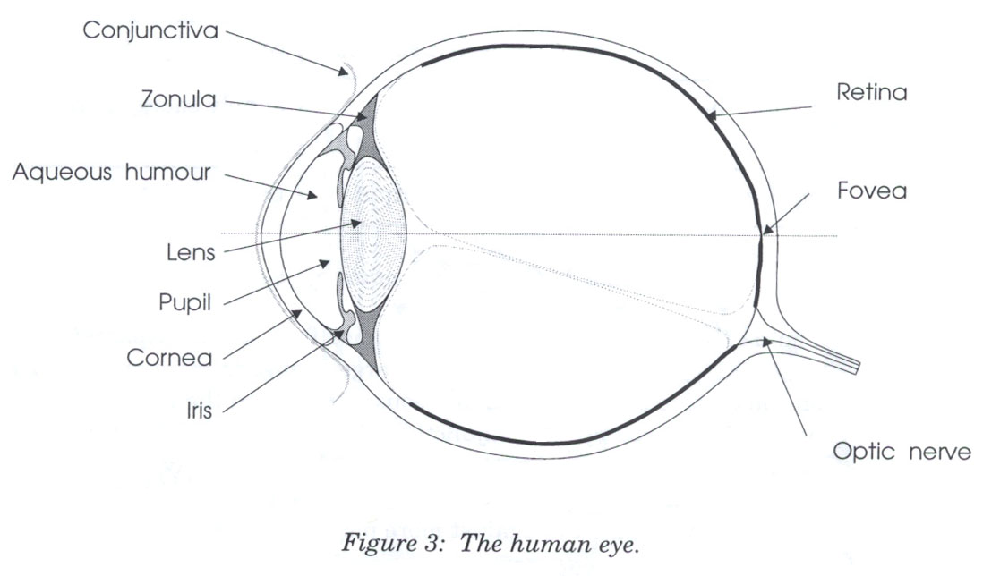
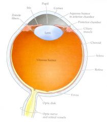
Parallel categories diagram in Python - Plotly Basic Parallel Categories Diagram with graph_objects¶ This example illustrates the hair color, eye color, and sex of a sample of 8 people. The dimension labels can be dragged horizontally to reorder the dimensions and the category rectangles can be dragged vertically to reorder the categories within a dimension. Eye Diagram With Labels and detailed description - BYJUS A brief description of the eye along with a well-labelled diagram is given below for reference. Well-Labelled Diagram of Eye The anterior chamber of the eye is the space between the cornea and the iris and is filled with a lubricating fluid, aqueous humour. The vascular layer of the eye, known as the choroid contains the connective tissue. BYJUS BYJUS Human eye - Wikipedia Each eye has seven extraocular muscles located in its orbit. Six of these muscles control the eye movements, the seventh controls the movement of the upper eyelid.The six muscles are four recti muscles – the lateral rectus, the medial rectus, the inferior rectus, and the superior rectus, and two oblique muscles the inferior oblique, and the superior oblique.
The Eye Diagram: What is it and why is it used? The eye diagram is used primarily to look at digital signals for the purpose of recognizing the effects of distortion and finding its source. To demonstrate using a Tektronix MDO3104 oscilloscope, we connect the AFG output on the back panel to an analog input channel on the front panel and press AFG so a sine wave displays. Then we press Acquire. brain parts labelled diagram labeled brain diagram left right labels anatomy parts sides drawing human difference science between functions structure results eye understanding memory ... brain diagram functions parts function different areas lobe labeled anatomy human frontal cerebellum lobes motor coordination example muscles helps shows. Cow's Eye Dissection - Eye ... PDF Eye Anatomy Handout - National Eye Institute of light entering the eye. Lens: The lens is a clear part of the eye behind the iris that helps to focus light, or an image, on the retina. Macula: The macula is the small, sensitive area of the retina that gives central vision. It is located in the center of the retina. Optic nerve: The optic nerve is the largest sensory nerve of the eye. Structure and Function of the Human Eye - ThoughtCo The main parts of the human eye are the cornea, iris, pupil, aqueous humor, lens, vitreous humor, retina, and optic nerve. Light enters the eye by passing through the transparent cornea and aqueous humor. The iris controls the size of the pupil, which is the opening that allows light to enter the lens. Light is focused by the lens and goes ...
Cell Membrane Structure Diagram | Cell Membrane Diagram | Biology | Pinterest | Models, Biology ...
Label the microscope — Science Learning Hub Jun 08, 2018 · Labels. Description. eye piece lens. The lens you look through – normally 10x or 15x magnification. coarse focus adjustment. Moves the lens up or down and adjusts focus. fine focus adjustment. Moves the lens in order to make very small adjustments to gain better focus. base. The bottom of the microscope used for stability. high-power objective
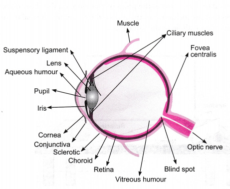
Diagram Human Eye Labeled - Aflam-Neeeak
Generate eye diagram - MATLAB eyediagram - MathWorks eyediagram(x,n) generates an eye diagram for signal x, plotting n samples in each trace. The labels on the horizontal axis of the diagram range between –1/2 and 1/2. The function assumes that the first value of the signal and every nth value thereafter, occur at integer times.

Draw a labeled diagram of human eye Write the functions of Cornea, Iris, Pupil, eye lens and ...
Labelled Diagram of Human Eye, Explanation and Function - VEDANTU The basic functions of Rods and Cones are conscious light perception, color differentiation and depth perception. The human eye is capable of distinguishing between about 10 million colors, and it can also detect a single photo. The human eye is a part of the sensory nervous system. Labeled Diagram of Human Eye
Slide Show: The Neural Control of Vision B-1
Structure And Function Of The Eye - Vision - MCAT Content The iris, which is conspicuous as the colored part of the eye, is a circular muscular ring lying between the lens and cornea that regulates the amount of light entering the eye. In conditions of high ambient light, the iris contracts, reducing the size of the pupil at its center. In conditions of low light, the iris relaxes and the pupil enlarges.

Eye diagram by Firkin | Human eye diagram, Diagram of the eye, Eye structure
eye diagram with labelling eye diagram with labelling Lacrimal Gland diagram - The Eye Si (gh)t we have 9 Pictures about Lacrimal Gland diagram - The Eye Si (gh)t like Internal Parts and Functions of the Eye | hubpages, Unlabeled Eye Diagram - ClipArt Best and also Unlabeled Eye Diagram - ClipArt Best. Here you go: Lacrimal Gland Diagram - The Eye Si (gh)t
Diagram Of The Eye Not Labeled - Diagram Media
Eye Anatomy: Parts of the Eye and How We See Behind the anterior chamber is the eye's iris (the colored part of the eye) and the dark hole in the middle called the pupil. Muscles in the iris dilate (widen) or constrict (narrow) the pupil to control the amount of light reaching the back of the eye. Directly behind the pupil sits the lens. The lens focuses light toward the back of the eye.
Eye Anatomy Diagramvector Illustration Stock Vector (Royalty Free) 485878195 - Shutterstock
eye diagram labeled brain function structure functions diagram lobes macmillan labelled showing. Cow Eye Dissection - YouTube . cow eye dissection. Label The Muscles Of The Eye - PurposeGames . purposegames. 3d Eye Model 32 Pcs Assembled Human Anatomy Model New 3D Structure Of . auge. Photoreceptor Cell ...

Diagram of the Eye
Human Eye Diagram, How The Eye Work -15 Amazing Facts of Eye First, light rays enter the eye through the cornea, the clear front "window" of the eye. The dome shaped cornea bends light to help the eye focus. From the cornea, the light passes through an opening called the pupil. The amount of light passing through is controlled by the iris, or the colored part of your eye.
Reading 15: Color
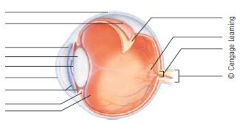
Labelling the eye — Science Learning Hub In this interactive, you can label parts of the human eye. Use your mouse or finger to hover over a box to highlight the part to be named. Drag and drop the text labels onto the boxes next to the eye diagram If you want to redo an answer, click on the box and the answer will go back to the top so you can move it to another box.
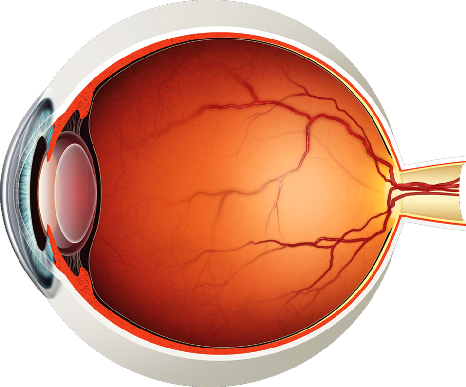
Diagram of the Eye - Lions Eye Institute
Parts of Stereo Microscope (Dissecting microscope) - Rs' Science If you would like to learn optical components of a compound microscope, please visit Compound Microscope Parts – Labeled Diagram and their Functions, and this article. How to use a stereo (dissecting) microscope. Follow these steps to put your stereo microscopes in work: 1. Set your microscope on a tabletop or other flat sturdy surface where ...

describe the function of eye and draw its labelled diagram - Brainly.in
Control Unit Installation and Operation Guide Please Read between any Eye QS control unit and any other power supply, including another GRAFIK Eye QS control unit. Refer to the QS Link Power Draw Units specification submittal (Lutron P/N 369405) for more information concerning PDUs. 1234 12 ABC 123456LN Example: Emergency lighting interface (maximum 1) Note: The GRAFIK Eye QS control unit
Label the parts of the eye: | bartleby
Parts Of The Eye Labeled Diagram Model And Their Function Parts of the eye-labeled diagram model are divided into three groups: the external outer layer, the middle layer, and the inner back layer. The outer layer is responsible for protecting the eye from environmental toxins and debris. The middle layer includes cells that allow light to enter and travel through the back layer to the retina.

Eye Diagram Without Labels | via Anatomy Pictures Gallery if… | Flickr
Labelling the eye — Science Learning Hub Labelling the eye Add to collection The human eye contains structures that allow it to perceive light, movement and colour differences. In this activity, students use online or paper resources to identity and label the main parts of the human eye. By the end of this activity, students should be able to: identify the main parts of the human eye
/GettyImages-695204442-b9320f82932c49bcac765167b95f4af6.jpg)
30 Parts Of The Eye With Label - Labels For Your Ideas
Eye Anatomy Diagram - EnchantedLearning.com Retina - light-sensitive tissue that lines the back of the eye. It contains millions of photoreceptors (rods and cones) that convert light rays into electrical impulses that are relayed to the brain via the optic nerve. Rods - cells the in the retina that sense brightness (they are photoreceptors). Night vision involves mostly rods (not cones).
Overview of Visual Systems Flashcards - Cram.com
PDF Parts of the Eye - National Eye Institute | National Eye Institute To understand eye problems, it helps to know the different parts that make up the eye and the functions of these parts. Here are descriptions of some of the main parts of the eye: ... Handout illustrating parts of the eye Keywords: parts of the eye, eye diagram, vitreous gel, iris, cornea, pupil, lens, optic nerve, macula, retina ...

Human Eye Diagram Labeled - Health Picture Reference | Eye care, Human eye, Human eye diagram
Eye anatomy: A closer look at the parts of the eye The iris of the eye functions like the diaphragm of a camera, controlling the amount of light reaching the back of the eye by automatically adjusting the size of the pupil (aperture). The eye's crystalline lens is located directly behind the pupil and further focuses light.

Medical Encyclopedia - Structure and Function: How the Eye Works | Structure and function ...
Create a Briliant Process Flow Diagram with Canva Process flow diagrams illustrate how a large complex process is broken down into smaller functions and how these fit together. As visual tools, they can help your team or organization see the bigger picture as well as where they fit into its entirety. Create a process flow any time you want to illustrate the stages of a process.
Post a Comment for "43 eye diagram with labels and functions"