40 picture of compound microscope with labels
microscope labeling worksheet microscope labeling parts label diagram compound biology worksheets labeled corner use worksheet cell microscopes quiz study test drawing science animal. What Is A Microscope And How It Works - 2017 bestmicroscopecentral.com. microscope parts biology diagram lab identification light functions microscopy compound lens drawing cartoon does ... Parts of a Compound Microscope and Their Functions - NotesHippo Compound microscope mechanical parts (Microscope Diagram: 2) include base or foot, pillar, arm, inclination joint, stage, clips, diaphragm, body tube, nose piece, coarse adjustment knob and fine adjustment knob. Base: It's the horseshoe-shaped base structure of microscope. All of the other components of the compound microscope are supported ...
Compound Microscope- Definition, Labeled Diagram, Principle, Parts, Uses In order to ascertain the total magnification when viewing an image with a compound light microscope, take the power of the objective lens which is at 4x, 10x or 40x and multiply it by the power of the eyepiece which is typically 10x. Therefore, a 10x eyepiece used with a 40X objective lens will produce a magnification of 400X.
Picture of compound microscope with labels
Ketoconazole cream vs. clotrimazole cream - MedicineNet A physical examination of the affected skin, evaluation of skin scrapings under the microscope, and culture tests can help doctors make the appropriate distinctions. A proper diagnosis is essential to successful treatment. Labeling the Parts of the Microscope | Microscope World Resources Labeling the Parts of the Microscope. This activity has been designed for use in homes and schools. Each microscope layout (both blank and the version with answers) are available as PDF downloads. You can view a more in-depth review of each part of the microscope here. #1 CGC | eBay Wij willen hier een beschrijving geven, maar de site die u nu bekijkt staat dit niet toe.
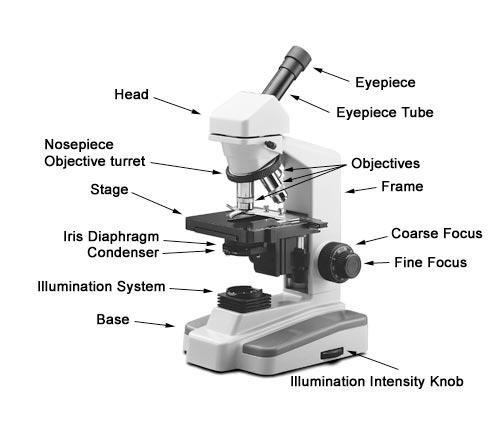
Picture of compound microscope with labels. Looking at the Structure of Cells in the Microscope ... This latter quantity is a measure of the width of the entry pupil of the microscope, scaled according to its distance from the object; the wider the microscope opens its eye, so to speak, the more sharply it can see . Under the best conditions, with violet light (wavelength = 0.4 μm) and a numerical aperture of 1.4, a limit of resolution of ... Labelled Diagram of Compound Microscope - Biology Discussion The below mentioned article provides a labelled diagram of compound microscope. Part # 1. The Stand: The stand is made up of a heavy foot which carries a curved inclinable limb or arm bearing the body tube. The foot is generally horse shoe-shaped structure (Fig. 2) which rests on table top or any other surface on which the microscope in kept. Compound Microscope: Definition, Diagram, Parts, Uses, Working ... - BYJUS A microscope with a high resolution and uses two sets of lenses providing a 2-dimensional image of the sample. The term compound refers to the usage of more than one lens in the microscope. Also, the compound microscope is one of the types of optical microscopes. The other type of optical microscope is a simple microscope. Microscope Diagram and Functions | Science fair projects, Microscope ... A Study of the Microscope and its Functions With a Labeled Diagram To better understand the structure and function of a microscope, we need to take a look at the labeled microscope diagrams of the compound and electron microscope. These diagrams clearly explain the functioning of the microscopes along with their respective parts. M mooketsi
Parts of the Microscope with Labeling (also Free Printouts) Parts of the Microscope with Labeling (also Free Printouts) By Editorial Team March 7, 2022 A microscope is one of the invaluable tools in the laboratory setting. It is used to observe things that cannot be seen by the naked eye. Table of Contents 1. Eyepiece 2. Body tube/Head 3. Turret/Nose piece 4. Objective lenses 5. Knobs (fine and coarse) 6. Compound Microscope with labels Stock Vector | Adobe Stock Download Compound Microscope with labels Stock Vector and explore similar vectors at Adobe Stock. Adobe Stock Photos Illustrations Vectors Videos Audio Templates Free Premium Editorial Fonts Parts of a microscope with functions and labeled diagram - Microbe Notes Head - This is also known as the body. It carries the optical parts in the upper part of the microscope. Base - It acts as microscopes support. It also carries microscopic illuminators. Arms - This is the part connecting the base and to the head and the eyepiece tube to the base of the microscope. Micro Module 1 Flashcards | Quizlet Different brands of light microscopes are slightly different but they usually contain the same basic components of a compound light microscope---ocular lenses in eyepieces, an arm, a revolving nosepiece with several objectives, a stage with some adjustment knobs to move the slide around on the stage, a condenser and iris diaphragm under the stage, two focus knobs, and a light …
Compound Microscope - Diagram (Parts labelled), Principle and Uses See: Labeled Diagram showing differences between compound and simple microscope parts Structural Components The three structural components include 1. Head This is the upper part of the microscope that houses the optical parts 2. Arm This part connects the head with the base and provides stability to the microscope. MSMEmart India - Indian Manufacturers, Suppliers, Buyers, … MSME Global mart is an Indian business to business (B2B) portal facilitating online marketing support to Indian micro, small, medium enterprises (MSMEs) Manufacturers, Suppliers and buyers through way of increased visibility, connecting buyers & suppliers, trade leads & keyword based unlimited tender alerts to grow their business. Microscope Labeled Pictures, Images and Stock Photos Browse 49 microscope labeled stock photos and images available, or start a new search to explore more stock photos and images. Newest results Fluorescent Imaging immunofluorescence of cancer cells growing... Microscope diagram vector illustration. Labeled zoom instrument... Microscope diagram vector illustration. What is Electron Microscopy? - UMASS Medical School Because the size of the raster at the specimen is much smaller than the viewing screen of the CRT, the final picture is a magnified image of the specimen. Appropriately equipped SEMs (with secondary, backscatter and X-ray detectors) can be used to study the topography and atomic composition of specimens, and also, for example, the surface distribution of immuno-labels.
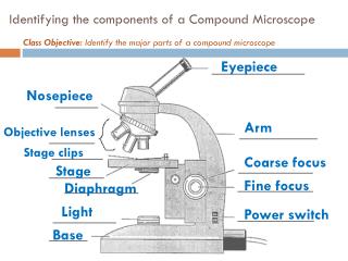
PPT - Identifying the components of a Compound Microscope PowerPoint Presentation - ID:2045335
Microscope picture label Flashcards | Quizlet Start studying Microscope picture label. Learn vocabulary, terms, and more with flashcards, games, and other study tools.

labels of a compound microscope microscope boxed - Top Label Maker
How to draw compound of Microscope easily - step by step I will show you " How to draw compound of microscope easily - step by step "Please watch carefully and try this okay.Thanks for watching.....#microscopedrawi...

microscope - Kids | Britannica Kids | Homework Help
Compound microscope Images, Stock Photos & Vectors - Shutterstock Compound microscope images 3,117 compound microscope stock photos, vectors, and illustrations are available royalty-free. See compound microscope stock video clips Image type Orientation People Artists Sort by Science College and University Insects and Spiders Jobs/Professions optical microscope scientist

Parts of a Microscope - HaleyMullmicroscopy
Compound Microscope Parts, Functions, and Labeled Diagram Compound Microscope Definitions for Labels. Eyepiece (ocular lens) with or without Pointer: The part that is looked through at the top of the compound microscope. Eyepieces typically have a magnification between 5x & 30x. Monocular or Binocular Head: Structural support that holds & connects the eyepieces to the objective lenses.

Clipart Panda - Free Clipart Images
Search - Defense Logistics Agency 17-11-2014 · This Site describes the products and services that DLA Logistics Information Service provides.
Print Microbiology Lab 2 (Microscopy, Gram stain, intro to enterotube II) flashcards | Easy ...
(PDF) Introduction to Microscopy - ResearchGate Nov 08, 2017 · • In compound microscope it will be i.e 10 X, f= 16 mm; 40 X, f= 4 mm; 100 X, f= 1.8 mm. • Image produced by objective lens falls on the eyepiece lens serve as objec t. • Image formed in the ...

Types of Microscope🔬| Simple-Compound-Special Microscope | - YouTube
The Parts of a Microscope (Labeled) Printable - TeacherVision The Parts of a Microscope (Labeled) Printable. Download. Add to Favorites. Share. This diagram labels and explains the function of each part of a microscope. Use this printable as a handout or transparency to help prepare students for working with laboratory equipment.
virtual microscope lab - YouTube
Lower Secondary Science LEARNER’S BOOK 8 - Issuu 22-02-2021 · Activity 1.5.1 Making a picture of blood You are going to make a picture of some blood, as it might look if you saw it through a microscope. Work …

View Product Photos
What is a Compound Microscope? - Study.com The body of the compound light microscope is the main part of the microscope, not to include the lights, focusing block, or stand of the microscope. The objective lenses and eyepiece are a part of ...

Microscope World Blog: Biological Microscope Magnifications
Micro Module 1 Flashcards | Quizlet Study with Quizlet and memorize flashcards containing terms like Move the terms into the correct empty boxes to complete the concept map., Drag the images and/or statements to their corresponding class to test your understanding of the main types of microbes., Drag the images or descriptions to their corresponding class to test your understanding of the cellular organization and relative size ...

shikha mahajan: Compound Microscope
What is Electron Microscopy? - UMASS Medical School Conventional scanning electron microscopy depends on the emission of secondary electrons from the surface of a specimen. Because of its great depth of focus, a scanning electron microscope is the EM analog of a stereo light microscope. It provides detailed images of the surfaces of cells and whole organisms that are not possible by TEM.

31 Compound Microscope With Label - Labels For Your Ideas
Laboratory Information System (LIS) - HEALTHCARE SERVICE … 06-09-2014 · Date First Published: September 6, 2014Date Last Revised: March 20, 2020 The name Laboratory Information System is more appropriate than say Pathology information system, since the Pathology department serves many functions, and systems should be designed to be functional rather than departmental. Clinical functions such as clinical microbiology …
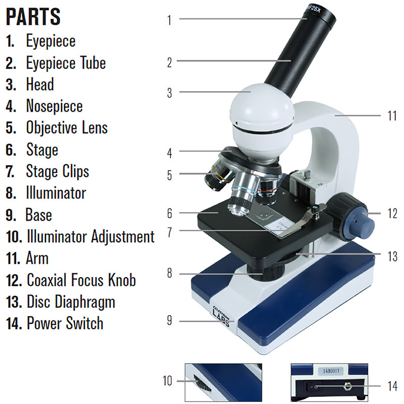
Celestron Labs Compound Microscope CM1000C
16 Parts of a Compound Microscope: Diagrams and Video Once you have an understanding of the parts of the microscope it will be much easier to navigate around and begin observing your specimen, which is the fun part! The 16 core parts of a compound microscope are: Head (Body) Arm. Base. Eyepiece. Eyepiece tube.
How to Use a Compound Microscope Basic Microscopy
(PDF) Introduction to Microscopy - ResearchGate 08-11-2017 · • In compound microscope it will be i.e 10 X, f= 16 mm; 40 X, f= 4 mm; 100 X, f= 1.8 mm. • Image produced by objective lens falls on the eyepiece lens serve as objec t. • …
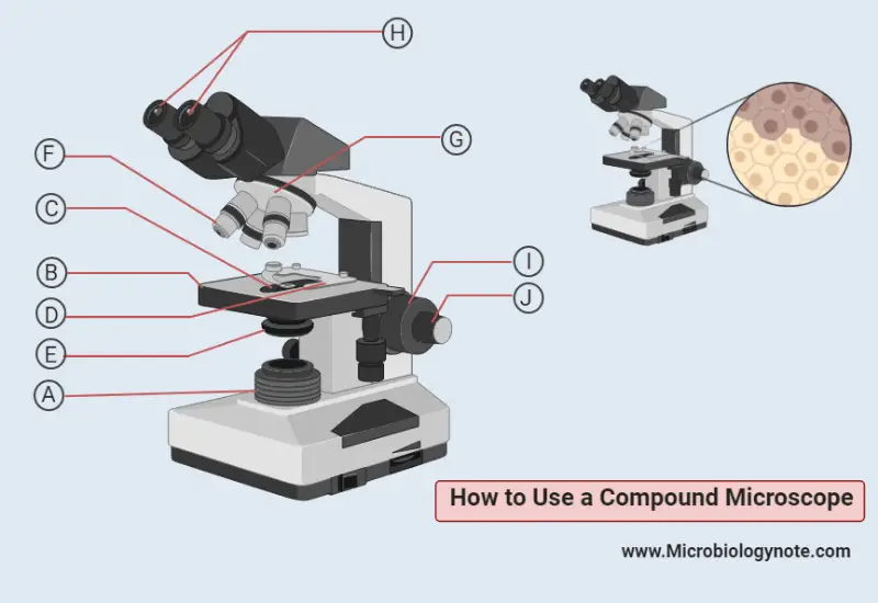
How to Use a Compound Microscope
A Study of the Microscope and its Functions With a Labeled Diagram ... These labeled microscope diagrams and the functions of its various parts, attempt to simplify the microscope for you. However, as the saying goes, 'practice makes perfect', here is a blank compound microscope diagram and blank electron microscope diagram to label. Download the diagrams and practice labeling the different parts of these ...

SCIENCE :: PHYSICS: OPTICS :: MAGNIFYING GLASS AND MICROSCOPES :: BINOCULAR MICROSCOPE image ...
Compound Microscope - Types, Parts, Diagram, Functions and Uses A compound microscope captures an inverted image of the specimen because every time the light passes through the lens, the image's direction is flipped. The image always ends up inverted from the original. So, if you move the sample to the left, it moves in the right direction. Image 18: A comparison image between a simple and compound microscope.

How To Use a Compound Microscope - YouTube
Compound Microscope Parts - Labeled Diagram and their Functions Basically, compound microscopes generate magnified images through an aligned pair of the objective lens and the ocular lens. In contrast, "simple microscopes" have only one convex lens and function more like glass magnifiers. [In this figure] Two "antique" microscopes played significant roles in the history of biology.
Post a Comment for "40 picture of compound microscope with labels"